Image Quality Factors: What Makes a Radiograph Diagnostic?
When evaluating radiographic images, it’s crucial to understand what makes them diagnostically useful. A high-quality radiograph should provide clear anatomical detail while minimising distortion and artefacts.
The four fundamental factors that influence image quality are contrast, density, resolution, and distortion. This article explores these aspects and how radiographers can optimise them.
Contrast: Differentiating Between Tissues
💡Definition: Contrast refers to the difference in shades of gray on a radiograph. A high-contrast image has distinct black-and-white regions, while a low-contrast image contains many shades of gray.
Factors Affecting Contrast:
- Kilovoltage Peak (kVp):
- Higher kVp results in lower contrast (more gray shades) due to greater penetration. More scatter will exit the patient and sometimes create unwanted fog around the anatomy.
- Lower kVp produces higher contrast with more distinct black-and-white separation. This may cause an abruption of density differences, causing subtle details to be missed or not imaged.
Therefore, it is important that we find a balance between the high and low kVp. Typically, for the healthcare setting, we choose a higher kVp, but we ensure it is not too high.
2. Subject Contrast:
- Differences in tissue density affect contrast. For example, bones (high density) absorb more X-rays than soft tissues (low density), leading to greater contrast.
3. Scattered Radiation:
- Scattered radiation reduces contrast by creating a blanket of unwanted exposure. Using a grid can help absorb scatter and improve image clarity.
📌The diagram below shows us how the difference between high and low contrast radiographs.
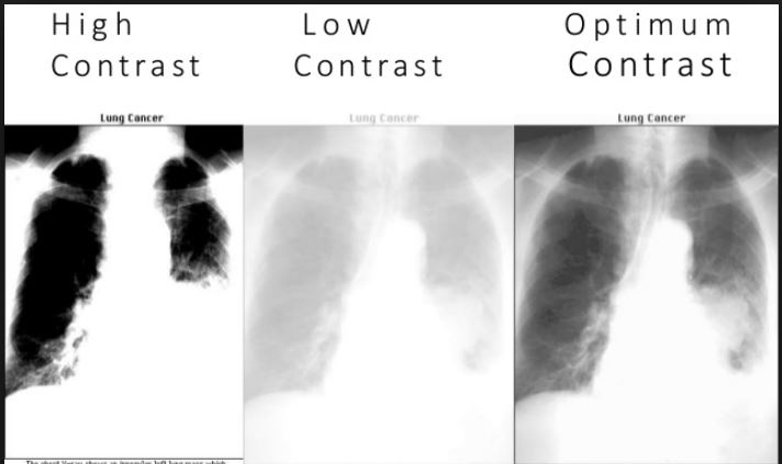
So high contrast is black and white, whereas low contrast is paler and greyer as they have a longer grayscale.
Density: The Overall Blackness of the Image
💡Definition: Radiographic density refers to the degree of blackening on the image. A well-balanced density ensures all anatomical structures are visible without being too dark or too light. In terms of image critiquing, we should be able to see the trabecular markings of the cortical bone nicely.
Factors Affecting Density:
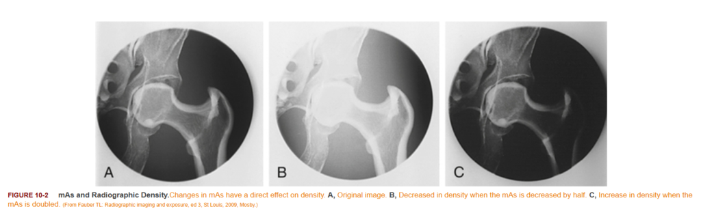
- Milliampere-Seconds (mAs) is the number of x-rays:
- Higher mAs increases density (darker image) by delivering more X-ray photons.
- Lower mAs decreases density (lighter image), which may obscure details.
- ↑ number of photons; ↑ number of interactions & image density; ↑ patient absorbed dose
- kVp Adjustments:
- Besides affecting contrast, increasing kVp also increases density due to higher X-ray penetration.
- Patient Thickness:
- Thicker body parts absorb more X-rays, requiring higher mAs or kVp to maintain adequate density, e.g. the abdomen.
📌We can comparing the different mAs settings on the hip joint below:
Resolution: The Sharpness of Detail
💡Definition: Resolution (also known as spatial resolution) describes how well small details can be distinguished on a radiograph. High-resolution images show clear structures with defined edges, while low-resolution images appear blurry.
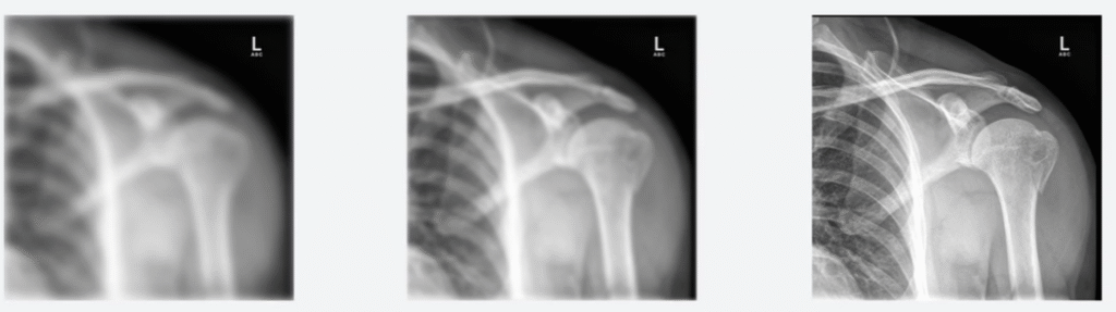
📌 We can see that the first image (low resolution) appears very blurry, and the edges are not sharp at all. The middle image has increased spatial resolution. The end image has the highest spatial resolution, where we can distinguish the fine details.
Image Souce – Clover Learning
Factors that can affecting Resolution:
- Focal Spot Size:
- A smaller focal spot improves resolution by producing finer X-ray beams.
- Motion Blur:
- Patient movement during exposure reduces resolution; using shorter exposure times (controlling the s from mAs) helps minimise motion artifacts.
- Receptor System:
- Digital radiography systems vary in pixel size and detector quality, which influence image sharpness.
Distortion: Maintaining True Object Shape
💡Definition: Distortion refers to the misrepresentation of the size or shape of anatomical structures on a radiograph.
Types of Distortion:
- Size Distortion:
- Occurs when an object appears larger due to increased object-to-image distance (OID).
- Shape Distortion:
- Caused by improper alignment of the X-ray beam, subject, or image receptor, leading to foreshortening or elongation.
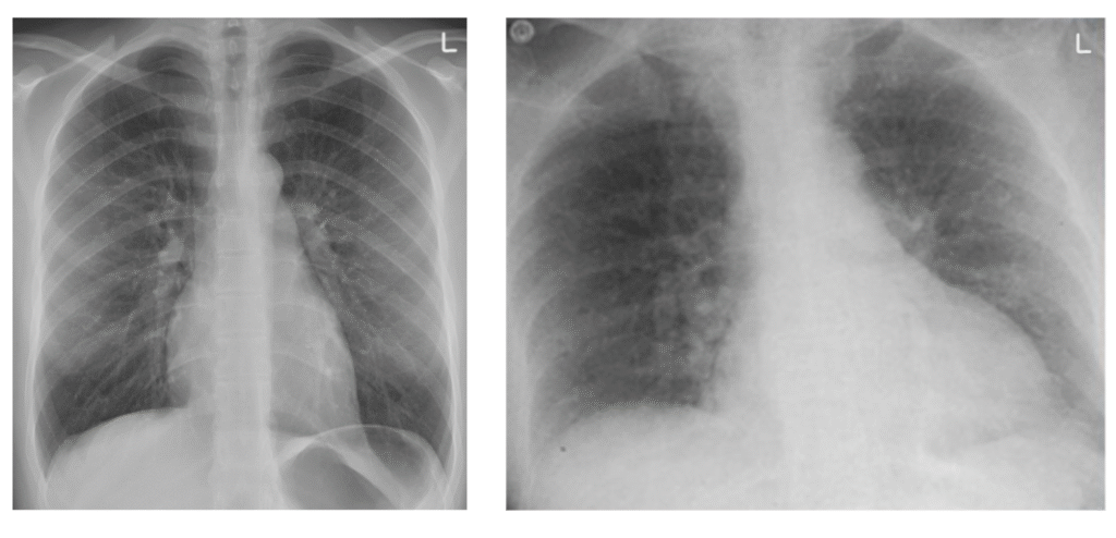
📌We can see below a standard chest xray on the left and on the right, we can observe the effects of shape distortion that has caused our anatomy to seem wider than in reality: